
Vulvar Anatomy: Understanding Your Own Body
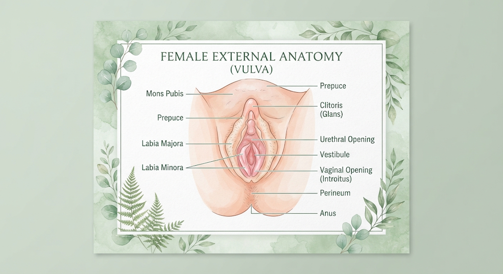
Most women were never taught the names or functions of their own external genitalia. School biology covered reproduction in the abstract, but rarely provided a clear map of the anatomy itself. Many women reach adulthood, and sometimes much further, without being able to accurately name what they are looking at, without knowing what normal variation looks like, or without feeling entitled to that knowledge in the first place. That gap is not trivial. It affects how confidently women can describe symptoms to healthcare providers, how quickly they notice changes, and how they feel about their own bodies. This article is a straightforward, clear guide to vulvar anatomy, written for every woman who deserved this information earlier.
The vulva is the correct term for the external female genitalia, including the labia majora, labia minora, clitoris, clitoral hood, urethral opening, vaginal opening, and the surrounding tissue. The vagina is the internal canal. Normal vulvar anatomy varies enormously between individuals, particularly in the size, shape, and colour of the labia and clitoris. Knowing your own anatomy matters because it allows you to notice changes, describe symptoms accurately, and seek help promptly when something is different.
Vulva Versus Vagina: Getting the Terms Right
The most pervasive anatomical error in everyday language is using “vagina” to refer to the entire female genital area. The vagina is specifically the muscular internal canal that connects the uterus to the outside of the body. It is internal.
The vulva is the collective term for all the external genitalia. Understanding this distinction matters beyond semantics. When a woman tells a doctor she has pain “down there,” or that something looks different, the ability to specify whether it is a symptom of the internal vaginal canal or the external vulvar structures helps direct the right examination and investigation. Using accurate terminology is a practical health tool, not pedantry.
The Structures of the Vulva
The labia majora are the outer folds of skin that run from the pubic mound down and around the vaginal opening. They contain fatty tissue and are covered externally by skin that typically has pubic hair. They serve as protective outer lips covering the more sensitive internal structures.
The labia minora are the inner folds of skin that sit inside the labia majora. They vary more dramatically between individuals than almost any other anatomical structure. In some women they are small and tucked inside the labia majora. In others they extend beyond the outer lips. They may be symmetrical or asymmetrical. They range in colour from pale pink to dark brown to a reddish-purple. All of these presentations are entirely normal. The labia minora have no standard appearance, and the images historically used in medical textbooks have been criticised for presenting a narrow, unrepresentative range.
The clitoris is considerably larger than most women are taught. The visible portion, called the glans clitoris, is the small, rounded structure located at the top of the vulva where the labia minora meet, partly covered by the clitoral hood. What is less widely known is that the clitoris extends internally, with two crura (legs) and two vestibular bulbs that extend along either side of the vaginal canal. The full internal structure of the clitoris was not accurately mapped in anatomical literature until relatively recently, and it remains absent from many medical textbooks. The clitoral glans is densely innervated with more than 10,000 nerve endings, making it the primary structure of sexual sensation in most women.
The clitoral hood is a fold of skin that covers and protects the glans clitoris, analogous in development to the male foreskin.
The urethral opening is the small opening through which urine exits the body. It sits below the clitoris and above the vaginal opening. It is not part of the reproductive system, but because of its proximity to the vaginal opening and the short length of the female urethra, it is relevant to understanding why urinary tract infections are more common in women.
The vaginal opening is the entrance to the internal vaginal canal. Around the vaginal opening, on either side, are the openings of the Bartholin glands, which produce small amounts of lubricating fluid. The Skene’s glands, sometimes called the female prostate, open near the urethral opening.
The perineum is the short stretch of tissue between the vaginal opening and the anus.
The Hymen: Clearing Up Persistent Myths
The hymen is a thin, membranous tissue that partially surrounds or covers the vaginal opening. It varies considerably in shape and size between individuals. Most hymens are not a complete seal but a partial crescent or ring of tissue with a natural opening.
The idea that the hymen is an indicator of virginity is medically inaccurate. Hymenal tissue varies so much between individuals and changes through normal activity, tampon use, and physical development that it cannot indicate sexual history. It also naturally changes over time and becomes less distinct after puberty and with age. This myth has caused significant harm and has no anatomical basis.
How Vulvar Anatomy Changes Through Life
Vulvar anatomy is not static. It changes meaningfully across a lifetime.
During puberty, the labia develop and darken, pubic hair appears, and the structures grow and mature. During the reproductive years, the labia minora and clitoris may respond to increased blood flow during arousal. Pregnancy and childbirth can alter the appearance and sensation of the perineum, labia, and vaginal opening. In perimenopause and postmenopause, reduced oestrogen leads to thinning and reduced elasticity of the vulvar and vaginal tissues, a condition sometimes called genitourinary syndrome of menopause (GSM), which can cause dryness, discomfort, and altered appearance.
Self-Examination: Why It Matters
Familiarity with your own vulvar anatomy is a genuine health tool. Women who know what their vulva normally looks and feels like are better placed to notice changes such as new lumps or skin changes, areas of persistent itching or soreness, changes in colour or texture of the skin, and unusual lesions. These can be signs of conditions including lichen sclerosus, vulvar dermatitis, or rarely, vulvar cancer, all of which are better treated when identified early.
Self-examination involves using a hand mirror in good light to look at the entire vulvar area. There is no particular technique required, only familiarity and the willingness to look. A gynaecology or primary care appointment is appropriate whenever you notice a change that is new, persistent, or uncertain.
FAQ
Is it normal for one labia minora to be larger than the other?
Yes. Labial asymmetry is extremely common. The two labia minora are independent structures and frequently differ in size, length, or shape. A review of anatomical studies shows that labia minora length ranges from less than one centimetre to more than five centimetres, and asymmetry between sides is the norm rather than the exception. Medical intervention is only warranted if there is significant discomfort, chafing during physical activity or sex, or difficulty with hygiene, not on the basis of appearance alone.
Can I use vulvar anatomy to identify health problems myself?
Familiarity with your own anatomy allows you to notice changes rather than diagnose conditions. Changes worth bringing to a healthcare provider include any new lump or raised area, persistent itching, changes in skin colour or texture (particularly whitening, thickening, or raw-looking skin), any sore that does not heal within a few weeks, and any area that bleeds without obvious cause. You do not need to know what these changes mean, only that they are different from your normal.
What is labial hypertrophy and when does it need treatment?
Labial hypertrophy refers to labia minora that are larger than average, often defined clinically as extending more than four to five centimetres from base to edge, though this cut-off is somewhat arbitrary. The vast majority of women with prominent labia minora have no health problems related to them. Where treatment is sought, it is most appropriately based on functional difficulty (discomfort, chafing, difficulty with hygiene) rather than cosmetic concern alone. The increase in labiaplasty procedures in recent years largely reflects cultural rather than medical drivers, and decisions about any surgical intervention should be made with full information and without external pressure.
Does the vulva need special cleaning products?
No. The internal vaginal canal is self-cleaning and should not have any products introduced into it. The external vulva, including the labia, should be washed with warm water. A gentle, unfragranced soap may be used on the outer labia majora and the surrounding skin if desired. Scented wipes, sprays, washes, or any product marketed as feminine hygiene for internal use disrupts the natural pH and microbiome and frequently causes more irritation than it prevents.
Knowing Your Body Is the Foundation of Advocating for It. Mayno Is Here for That.
The more clearly you understand your own anatomy, the more confidently you can describe what has changed, ask the right questions, and recognise when something needs attention. That knowledge is not a luxury. It is a health tool every woman deserves to have. Mayno is here to give it to you.
Visit mayno.health for women’s health guidance that takes your body seriously.
This article is for informational purposes only and does not constitute medical advice. If you have concerns about vulvar or vaginal health, please consult a qualified healthcare professional. Mayno Health does not replace clinical care.
Disclaimer: This article is for informational purposes only and does not constitute medical advice. Always consult a qualified healthcare professional.